Before learning about annelids, I had no idea that earthworms even had organs, so the dissection was also beneficial in improving my understanding of the living world around me. I learned that earthworms do in fact have a nervous system and even a primitive brain composed of ganglia as well as a ventral nerve cord that runs the length of their body. Additionally, I learned that worms in fact breathe through their skin and that that is why they are constantly moist. Earthworms also have two blood vessels and five hearts. Had my partner and I not punctured the crop and gizzard of the worm, we would have been able to see more of these fascinating things, but at least we now know that surgeon would not be a good professional choice.
None of these systems are apparent from the outside, and it is interesting to note that even the simplest and most overlooked animals are quite complex beings on the inside.


The process of digestion in an earthworm necessitates the involvement and cooperation of many different organs. Food is ingested by the earthworm and then travels to the pharynx which pumps a mixture of food and soil into the esophagus. The food-soil mixture is then transported to the crop for storage and continues on to the gizzard where it is ground up into small pieces for more effective digestion by the intestine. Any undigested materials pass through the worm's intestine out through the anus in solid form.


The two large nerves that pass around the gut of the worm and connect the brain with a pair of ganglia are also attached to the ventral nerve cord that runs the length of the worm's body. These parts, the ganglia, nerves and ventral nerve cord make up the nervous system and the ganglia above the pharynx serve as the worm's brain.

The intestine and nephridia are included in the worm's excretory system. They both remove waste products from the body.

The easiest way to find out if an earthworm eats soil is to examine its excrement or castings. Because the soil is indigestible, it will come out in the excrement as a grainy substance.

Four pairs of earthworm setae. The earthworm's setae are very useful tools for the worm as its habitat is soil and sediment. It must burrow through the soil and sediment, and this is difficult without something like setae to anchor them in the soil. Setae are especially useful as a defense mechanism against animals which try to pull the worm up from the soil.


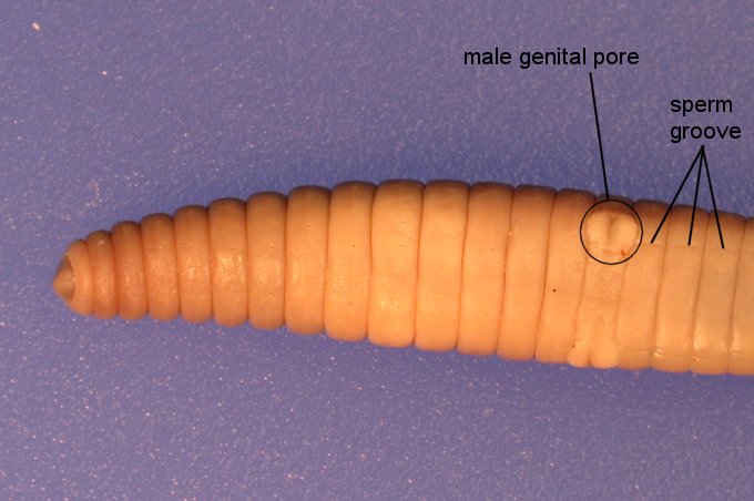
NOTE: These pictures are from google. I did have some pictures on my phone of the actual dissection, but I was unable to upload them.
Your google picts are good. You chose ones that showed each structure nice and clear.
ReplyDeletePurpose-4/4
Connection to class-4/4
Personal Reflection-4/4
Conventions-4/4
Requirements-9/9
25/25