Friday, January 25, 2013

Rat Dissection

   Last Thursday and Friday, our class dissected an infamous mammal. It carried the bubonic plague and still carries rabies. For some people, it is a terrifying creature, especially when beheld in one's kitchen. Ah, the rat; such a lovely creature. Rats definitely get a bad rap, but they're really not so bad. They're mammals trying to survive, just like ourselves.

  The dissection of the rat was very different from our previous dissections because the rat is a vertebrate. This means that it possesses a backbone, and is therefore far more advanced than an invertebrate. The rat is also a mammal, a member of our own phylum. Mammals are endothermic, hairy, air breathing and live bearing animals. Their most important characteristic and also the reason for their name, is that they possess mammary glands with which to nurse their young. Only female mammals can possess these mammary glands. Unfortunately, as we had a male rat, we were unable to observe them. However, the things we were able to observe (such as digestive and circulatory organs) proved very interesting, albeit slightly nauseating!

   In dissecting the rat, we come a little bit closer to understanding the inner workings of ourselves and the creatures around us. It becomes apparent, when one studies animals for a period of time, that some characteristics are common to many animals. In recognizing these common characteristics, it becomes easier to identify them in even the most radically different organisms. The gizzard of the earthworm and that of the bird, although they are completely different organisms from entirely different phyla, perform essentially the same function. In dissecting organisms, and not just the rat, we gain a greater understanding and appreciation for the  entire spectacular living world around us. It may not be the most pleasant of experiences, but as a teaching tool, it proves invaluable.


  1. Hands are the best tools in dissection because one can be more precise and delicate with the specimen than is possible using instruments. Additionally, it is beneficial to one's learning to be able to feel the texture, structure and stiffness of each organ.

  2. The purpose of having many different labels and titles for a dissection is to create a language with which to describe certain organs and be able to identify them using the names given.

  3. The tail differs from the rest of the rat's body in that it has far less hair and has an almost scaly appearance.
  4. The vibrissae on the rat act as sensory structures, allowing the rat to sense location, size, texture
    and other details on objects.
  5. Bilateral symmetry is a type of biological symmetry in which the organism is separated into two halves down the middle of its body, and each half is a mirror image of the other half.
  6. The sphincter of the rat is a ring shaped muscle, and it is shaped this way so that it can open and close the valves between various organs.
  7. The reason for the difference in size between the large and small intestine has to do with the matter that it handles. As the small intestine handles freshly digested food that is soft, there does not need to be too much room for food to pass through. The large intestine deals with food that has been digested well and is beginning to solidify and form feces. Therefore, less room is needed in the small intestine than in the large intestine because fecal matter takes up more space.
  8. The function of the liver in the rat is to digest fats, process nutrients, produce urea and cholesterol, to store vitamins and minerals and to filter the blood.
  9. The duodenum acquired its name from the Latin equivalent that means 12 finger widths (about 20 cm.)
  10. The appendix in rats in known as the cecum and is used for digesting high fibre plant materials containing cellulose. This digestion is done by bacteria in the cecum that release enzymes to digest the cellulose.
  11. The function of the membrane covering the walls of the body cavities and organs is to secrete a lubricating fluid that reduces friction from muscle movement.
  12. The function of the spleen in the rat is to clean the blood by destroying old blood cells and producing new ones.
  13. The function of the diaphragm in the rat is to expand and contract the thoracic cavity. This allows the lungs to expand to take in oxygen, and then deflate to release carbon dioxide.
  14. There are certain features that differentiate the atria from the ventricles. The smaller atria have thinner muscle tissue than the lower ventricles. Also, the atria pumps blood to the ventricle, and the ventricle pumps blood to the rest of the body. It is larger because it has a more complex function to perform than the atria.
  15. The left ventricle wall is thicker than the right ventricle wall because it needs to pump blood to the entire body whereas the right ventricle only needs to pump blood to the lungs to be oxygenated. Pumping blood to the entire body requires more muscle, so the left ventricle is thicker.
  16. Although the differences between the male and female reproductive systems of the rat are more pronounced, there are certain similarities between them as well. They both produce gametes, possess two adrenal glands, two kidneys, a urinary bladder, a urethra and an anus.
  17. The function of the kidneys in the rat are to excrete nitrogenous wastes in the form of urine and to regulate the water balance of the animal.
  18. The Thyroid, Thymus and Adrenal glands belong to the Endocrine system of the rat. The Thyroid gland controls how quickly the body uses energy, makes proteins and controls how sensitive the body is to other hormones. The Thymus gland functions in the development of the immune system by releasing hormones that stimulate the production of T-Cells. The Adrenal glands release hormones in response to stress, release androgens, and are involved in regulating the concentration of blood.




Wednesday, December 19, 2012

Inverterbrate Project

Hummingbird Clearwing Moth 
Hemaris Thysbe


The Hummingbird Clearwing Moth is one of nature's very best examples of mimicry and delicate beauty. Like the hummingbird, it hovers in the air to sip nectar from sweet flowers, beating its wings at rapid speeds so fast that they seem a blur. Although these moths share many similarites with the Hummingbird, that is not why they are called Hummingbird Moths. It is because, when sighted, they are often mistaken for hummingbirds themselves!


Phylum: Arthropoda
Class: Insecta 

Essential Functions

1) Body Structure
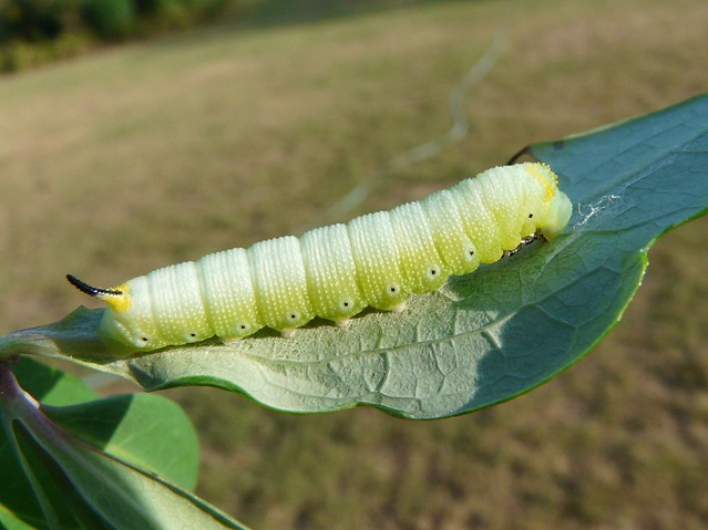
Adult Hummingbird Clearwing Moths grow to about 2 inches in length with wings that span 1-2 inches. Their thorax is olive in colour dorsally, and yellow ventrally, with a burgundy coloured abdomen ending in a fan-like tail of setae. The moth has one pair of compound eyes, one pair of strongly clubbed antennae, six legs and a proboscis. Their most interesting physical feature, as denoted by their name, is their clear wings. Their wings are scaled like most moths when they first leave their cocoon, but once they have taken first flight, the scales fall off and leave behind beautiful clear wings rimmed with orange-burgundy borders and veins. Hummingbird Clearwing larvae are most commonly green, brown and gray, but other colours do exist.


2) Digestion/Feeding
Adult Hummingbird Moths snack on the nectar of their favourite flowers. These include Japanese Honeysuckle, Beebalm, Red Clover, Lilac, Phlox, Snowberry, Cranberry and Blueberry plants and Thistles. Their larvae enjoy the leaves of these same plants. An adult moth uses a specialized mouthpart, a long, thin, needle like proboscis to suck up the nectar.

Like all moths, the Hummingbird Clearwing Moth's digestive system consists of a foregut, hindgut and midgut. After nectar is sucked up through the proboscis, it is passed into the esophagus and then stored in the crop for a short time. After this it continues on to the gizzard and then through the valve to the midgut and to the stomach where main digestion and absoption of nutrients takes place. The hindgut will then absorb the  water and nutrients from the nectar that the midgut did not and pass the waste out through the anus.

3) Circulation
 Hummingbird Clearwing Moths have an open circulatory system where blood is passed through blood vessels and body cavities called sinuses. Moth blood is actually green due to the fact that it is not oxidized when carried through the dorsal vessel. The moth has one heart that pumps blood to the rest of the body.

4) Respiration
Hummingbird Clearwing Moths breathe through thoracic and abdominal spiracles connected to tracheal tubes that diffuse oxygen to all body tissues and diffuse carbon dioxide out of the body through the spiracles.

5) Excretion
Moths excrete food wastes out through the anus. They also possess a set of Malpighian tubules which eliminate nitrogenous wastes from the blood, passing them out through the anus along with food wastes.

6) Movement
Hummingbird moths have a very unique way of moving unlike any other moth in their order. Their wings are made of many thin layers of chitin and they beat them very rapidly to hover in the air. Their wings can reach speeds of 30 beats per second and 30 miles per hour!


7) Reproduction
Hummingbird Clearwing Moth reproduction is internal and the moth breeds an average of 2-3 times per years. The moths detect mates by smell and sight, and once found, have been known to engage in a courtship chase in which the male and female fly through the air spiralling upward or flying low to the ground. Once the female's eggs are fertilized, she will lay them on a host plant. A host plant is one that the hatched larvae can feed upon (leaves) and are often the same plants it feeds on in adulthood. (nectar) The female Hummingbird Moth can lay up to 200 eggs, each on a different host plant! The eggs hatch into larvae within 6-8 days and after a while undergo complete metamorphosis, spinning a loose,glossy cocoon from which it will emerge an adult Hummingbird Clearwing Moth.


Other Important Information


  • Hummingbird Clearwing Moths are bilaterally symmetrical.
  • They have 3 germ layers: the mesoderm,(a middle layer of cells that forms
    the muscles and interior organs) endoderm, (cells on inside of gastrula that become the lining of the gut) and ectoderm. (cells on outside of gastrula that become the body covering). 
  •  Other organsims found within the same phyla as Hummingbird Moths (Arthropoda) include beetles, centipedes, spiders, butterflies, grasshoppers and many, many more.
  • Hummingbird Moths are protostomes. In protostome development, the embryo forms a dent on one side called the blastopore which deepens to become the archenteron which is the first growth of the gut. The original dent becomes the anus of the moth and the gut tunnels through the embryo to form the mouth of the moth.

Ecological Significance


  • Hummingbird Moths are important pollinators and aid in seed production.
  • They are food for a variety of wildlife including other insects, spiders, frogs, toads, lizards, shrews, hedgehogs and birds. That is, IF they can catch them!
  • Hummingbird Moth caterpillars eat certain plants' leaves, causing them to evolve characteristics which make them less appealing to the larvae. An example might be developing bad tasting chemicals on the surface of its leaves that deter the animals from eating them.
  • Hummingbird Clearwing Moths are useful as an indicator species as they are so abundant but are also quite sensitive to environmental change. 

Interesting Facts 
  • Hummingbird Clearwing Moths are seen on clear, sunny days unlike most moths because, like humans, they are diurnal and sleep at night.
  • The Hummingbird Moth mimics the Hummingbird in order to trick predators into staying away from it.
  • It lives in Alaska, the Northwest Territories, BC, Oregon, Maine, Newfoundland, Florida and Texas.  
  • Hummingbird Moths are known for being quite comfortable around humans and some will even land on you if you have some tasty nectar to offer!

 So why do I love Hummingbird Clearwing Moths? Because they defy our expectations with their fascinating mimicry and are adorable little creatures.


Monday, December 10, 2012

Squid Dissection

Last Friday our Biology class dissected a mollusk, one that is abundant in most marine environments, and is even one of the main characters on the popular kid's show, Spongebob Squarepants. We dissected Squidward, or at least someone related to him; a fifth cousin perhaps?

A squid is a cephalopod. In Latin this means "head foot". It sounds a little strange, but the name is actually quite logical. A squid really is just a head attached to some feet! Octopi are another well known species of this phylum. The two other types of mollusks that can be seen are known as Gastropods and Bivalves. One of the most common Gastropods in existence is the garden snail. Gastropods are "stomach feet", as their stomach is literally just above their feet inside their bodies. The next species in the phylum Molluska are the Bivalves. Their name means "two shell" as they possess two shells that can be tightly closed with strong muscles. Many of our most delectable seafood items, like clams and oysters are bivalves. It is interesting to note that although these forms are radically different from each other phenotypically, they all share specially evolved shells and feet. In fact, this is how they are classified. The squid, for example, has hardly any shell externally, but has a small plastic-like tube inside of it called the pen which is what is left of the squid's shell after evolution.

The squid has a most interesting internal make-up. Its insides are quite jelly like, save for the hard pen, and its ink sac looks quite a bit like a small minnow that has not yet been digested by the squid. It has a closed circulatory system rather than an open one like the bivalves as an open system is not conducive to free swimming and active animals such as squids. They possess advanced vision and brains and will secrete an ink cloud when threatened. It is difficult to explain the various features of the squid,  and how its internal organs function. Dissecting the squid allows us a first hand look inside its body, a real life explanation of its complex inner workings. Squids are fascinating   creatures and there is so much more to them than one sees as they glance into the plastic case at the T and T supermarket, and that is why dissection is so helpful to one's understanding of internal biology.


Our squid had 8 arms and 2 tentacles.

 The squid uses arms and tentacles for different purposes. Tentacles serve to help the squid grab prey and hold on to it and also play a role in mating whereas the arms are used to aid in feeding and grasping prey once it is in close proximity to the beak. Suckers on the arms are bigger than those on the tentacles.

 Forgot to take a picture for this one. To move, the squid draws water up into its mantle cavity and then expels it through the funnel creating jet propulsion. The squid can move either forward or backward.

Our squid's eye. Its eyes and tentacles are both adaptations for its predatory life. Its light-sensitive eyes allow it to see prey even in the darkness of the depths of the ocean, and its tentacles allow it to catch prey by surprise, snatching it at high speeds from far away.

Two traits that the squid shares with other mollusks are the presence of a mantle and of a visceral mass. Although all mollusks also have a foot, it is highly specialized and evolved to each mollusk's needs and thus looks different for every species.

The squid has 2 pairs of gills, one on each side.

The ink sac empties into the siphon and out of the squid's body into the surrounding water. Its function is to allow the squid camouflage in a dangerous situation so that it can quickly swim away from a predator.

This is the pen of our squid. The function of the pen is to give the squid structural support. It is considered a vestigial structure, so the squid's functions would most likely not be impaired were it not to have a pen.

As squids do possess a full digestive system, wastes exit the squid through the anus.


Monday, December 3, 2012

Earthworm Dissection

Last Friday, we dissected an earthworm in class. Although slightly icky, dissecting the earthworm allowed us to see first-hand the various parts of an annelid's nervous, respiratory and digestive system. The purpose of the dissection was also to improve our understanding of an annelid's internal organs so that we could identify them based on shape and function. 

Before learning about annelids, I had no idea that earthworms even had organs, so the dissection was also beneficial in improving my understanding of the living world around me. I learned that earthworms do in fact have a nervous system and even a primitive brain composed of ganglia as well as a ventral nerve cord that runs the length of their body. Additionally, I learned that worms in fact breathe through their skin and that that is why they are constantly moist. Earthworms also have two blood vessels and five hearts. Had my partner and I not punctured the crop and gizzard of the worm, we would have been able to see more of these fascinating things, but at least we now know that surgeon would not be a good professional choice.

None of these systems are apparent from the outside, and it is interesting to note that even the simplest and most overlooked animals are quite complex beings on the inside.

The name of the pumping organs in an earthworm are the aortic arches.

The process of digestion in an earthworm necessitates the involvement and cooperation of many different organs. Food is ingested by the earthworm and then travels to the pharynx which pumps a mixture of food and soil into the esophagus. The food-soil mixture is then transported to the crop for storage and continues on to the gizzard where it is ground up into small pieces for more effective digestion by the intestine. Any undigested materials pass through the worm's intestine out through the anus in solid form.

The two large nerves that pass around the gut of the worm and connect the brain with a pair of ganglia are also attached to the ventral nerve cord that runs the length of the worm's body. These parts, the ganglia, nerves and ventral nerve cord make up the nervous system and the ganglia above the pharynx serve as the worm's brain.

The intestine and nephridia are included in the worm's excretory system. They both remove waste products from the body. 

The easiest way to find out if an earthworm eats soil is to examine its excrement or castings. Because the soil is indigestible, it will come out in the excrement as a grainy substance.

http://biology.clc.uc.edu/graphics/bio113/worm/WormSetae.jpg
Four pairs of earthworm setae. The earthworm's setae are very useful tools for the worm as its habitat is soil and sediment. It must burrow through the soil and sediment, and this is difficult without something like setae to anchor them in the soil. Setae are especially useful as a defense mechanism against animals which try to pull the worm up from the soil.

The earthworm's digestive system is adapted for extracting relatively small amounts of food from large amounts of ingested soil. This is possible due to the gizzard of the worm which grinds up food particles into smaller pieces and then passes them on to the intestines where food is absorbed and soil is excreted through the anus. 


If we dissected the remainder of the worm to the posterior end, we would have observed the continuation of the intestine, the ventral nerve cord, and at the very end, the anus.

http://faculty.gvsu.edu/triert/biolab/bio111/erthwrm/eworm3c.jpg
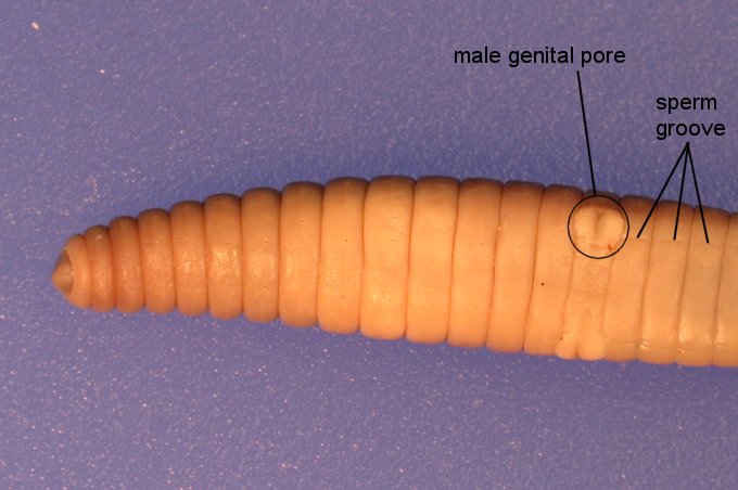
The earthworm's reproductive organs have certain roles and functions in order to carry out reproduction successfully. Because earthworms are hermaphrodites, they produce both sperm and eggs. Eggs are produced in the ovaries and sperm are produced in the testes. During reproduction, two worms connect at the clitellum and exchange sperm through their male genital pores. After copulation has occurred, the clitellum secretes a mucus-like cocoon which the worm slips out of, its eggs and the other worm's sperm being injected inside the cocoon through the female and male genital pores as the worm escapes it. Once the worm has left the cocoon, its ends close and embryonic worms begin developing.


NOTE: These pictures are from google. I did have some pictures on my phone of the actual dissection, but I was unable to upload them. 


Zoology Webquest

Porifera

An Azure Vase Sponge.
Genus: Callyspongia
Species: Callyspongia plicifera
The Azure Vase Sponge's colouring ranges from pink to purple and fluorescent light blue.

A Barrel Sponge.
Genus: Xestospongia
Species: Xestospongia muta
Barrel Sponges are known as the "Redwoods of the Reef" due to their 2000 year life span, size and colouring.
A Row Pore Rope Sponge.
Genus: Aplysina
Species: Aplysina cauliformis
This sponge inhabits lagoons and deep sloping reefs.


Cnidaria

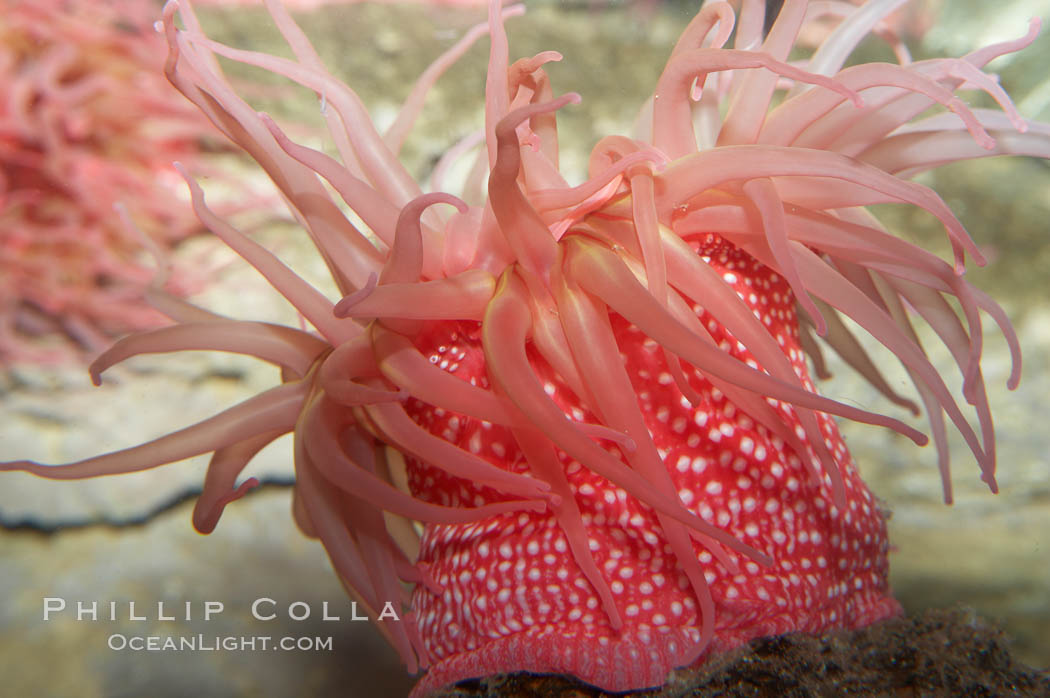
The Tealia Anemone.
Genus: Urticina
Species: Urticina Crassicornis
The Tealia Anemone may feed on crabs, sea urchins, mussels, gastropods, chitons, barnacles, fish, and sometimes sea stars and stranded jellies.

The Lion's Mane Jellyfish.
Genus: Cyanea
Species: Cyanea capillata
The Lion's Mane Jellyfish is the largest known jellyfish. It lives in the cold water of the Arctic, the northern Atlantic, and northern Pacific Oceans.


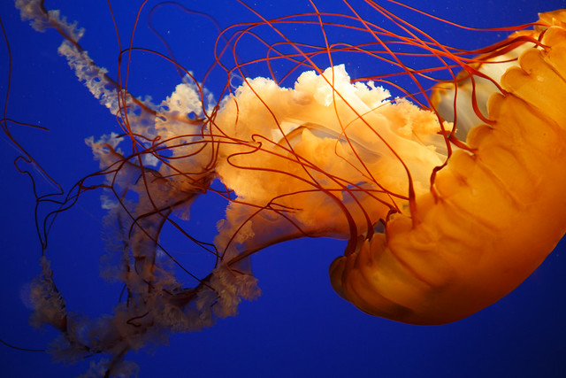
A Pacific Sea Nettle.
Genus: Cryasaora
Species: Chrysaora fuscescens
Pacific Sea Nettles live in the East Pacific Ocean from Canada to Mexico.


Platyhelminthes

The Persian Carpet Flatworm.
Genus: Pseudobiceros
Species: Pseudobiceros bedfordi
The Persian Carpet Flatworm eats ascidians and other crustaceans.

The Fuschia Flatworm
Genus: Pseudoceros
Species: ferrugineus 
The Fuschia flatworm can reach a length of about 18-48 mm.

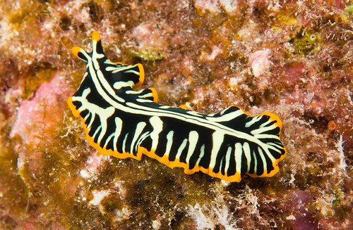
The Tiger Flatworm
Genus: Maritigrella
Species: Maritigrella crozierae
 Maritigrella crozierae is common in the Indian River Lagoon where it feeds exclusively on the mangrove tunicate, Ecteinascidia Turbinata.


Tuesday, November 27, 2012

Aquarium Trip

     Last Thursday, our Biology class went to the Vancouver Aquarium for the day. We were finally able to put a face to the names of the marine animals we learned about that past week. One's teacher can describe in great elaborate detail the movement of a jellyfish or the size of a beluga, but words cannot accurately illustrate these wonders. And so, it is necessary to see these amazing creatures with our own eyes to truly understand their fascinating lives. Additionally, one can learn many things one never knew from simply reading the descriptions beside the tanks.

       It feels good to glance in at the jellyfish and be able to identify their various parts and the stage of life that they are in; to touch the anenome and feel its sticky quality, knowing that it is in fact trying to sting you! In my opinion, this type of information will be the most valuable that I will learn in school, because it directly relates to the living world around me. In my opinion, there is far more satisfaction in being able to explain to someone the life cycle of a sponge, for example, than to be able to calculate the height of some building with trigonometry. Biology is the study of life, and what is more important or relevant than life itself?

      I enjoyed visiting the aquarium for the spectacular learning experience and just maybe the adorable sea otters were a little plus. :)

Here are some photos I took at the aquarium:

These are clown fish and sea anemones. Sea anemones will normally sting fish which are unlucky enough to come close to their tentacles and then digest them. However, the clown fish is immune to its stings and actually shares in a mutual symbiotic relationship with it. The clown fish is protected from some of its enemies by the anemone's stinging tentacles and the anemone, in return, is protected by the clown fish from other fish which would normally eat its tentacles.

This is a sea otter. They spend a lot of time cleaning their coats because it helps them to remain waterproof and insulated against the cold.

Before 1990, sea otters could be found from Russia to Mexico and in the waters of the North Pacific. They were reintroduced to Washington and Oregon in 1970 and it is now estimated that 500 sea otters exist in Washington today.


Next to the sea otters' tank were the green sea anemones.


This is Horn Coral. I picked it as the marine invertebrate that I found most interesting. It is actually a very aggressive coral and will kill most other corals near to it. This coral's colour is, interestingly enough, fluorescent green.

These are supposed to be the belugas but the picture quality is pretty awful. Their scientific name is Delphinapterus leucas. There are two belugas, named Aurora and Qila, at the aquarium.

This is a spotted seahorse. The movement of a seahorse is most interesting. It floats delicately in the water, its side fins beating furiously, much like a hummingbird's wings.

An open brain coral.

A starry flounder. It has a couple of quite interesting adaptations. First, it has pigment cells which allow it to change its colouring to suit its environment. Second, it undergoes a metamorphosis in which both eyes migrate to one side of the fish, the dorsal side. Most Starry Flounders are left eyed.


Moon jellyfish. They move by pushing water out of their bell, propelling themselves forward in a pulsing motion. Moon jellies have a very unique digestive adaptation. Food collects on the surface of the moon jelly, where it is then captured by the jellyfish's mucus and then passed on to the lappets. The food is then carried to eight different canals which all lead to the stomach.


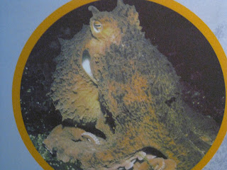
Unfortunately we were unable to find the Pacific Octopus, but above is a picture of what it looks like. The white suction cups underneath its eight arms serve to help it cling to things, grasp prey and other objects. The Pacific Octopus is known as one of nature's greatest predators because it is so large yet it is incredibly stealthy, and it will eat practically anything it can catch, including birds!


Steller sea lions eat many different kinds of fishes, including pacific herring, pollock, salmon, cod, and rockfishes. They can also eat octopi  and squids!

The caiman crocodile. This one was born in Fort Worth, Texas, before being transported to the Vancouver Aquarium by the National Zoological Park in Washington, DC. The colour of the crocodile is a muddy gray. This colouring would be beneficial in capturing prey as it would blend well into rocks, water and mud.
This is the Arapaima, the largest fish in the aquarium.

This is the red hook silver dollar. It is a vegetarian fish that can eat all of the plants in an aquarium if one is not careful!

A piranha. They pose the biggest threat to humans when we are fishing for them and removing the fishhooks from their mouths as they can bite one quite badly.

A shark. They have no bones. Instead, sharks possess cartilage, which is a strong, flexible material that is found all throughout their bodies.

A fish found in the shark tank. Sea turtles were also present.
This is the giant red sea urchin, also known as Strongylocentrotus franciscanus. They often lodge themselves into cracks and crevices to prevent themselves from being swept up in the ocean currents.

An archer fish. It feeds in a very interesting way. The archer fish will swim directly beneath an insect above the water surface, then stick its snout through the water and shoot the insect with a powerful stream of water. If the archer fish is successful, the insect falls into the water, right next to the fish and it is devoured.


This sea turtle's name is Schoona. She is a Hawksbill sea turtle.